Ph.D. Student Yun Shang Publishes in Magnetic Resonance in Medicine
An article published in the renowned scientific journal Magnetic Resonance in Medicine offers a new perspective on the long-standing issue of magnetic field inhomogeneity in the human heart during MRI scanning. This study revealed high-detailed information about the magnetic field variations in the human heart throughout the entire cardiac cycle, allowing clinicians and researchers to optimize MRI scans to achieve more accurate diagnosis in cardiovascular diseases.
The paper titled “High resolution simulation and measurement demonstrate oscillatory spatiotemporal B0 fluctuations across the human cardiac cycle” (https://pubmed.ncbi.nlm.nih.gov/37598417/) presents the culmination of years effort among a multidisciplinary team of cardiologist and engineers, led by Yun Shang, a Ph.D. student in the MR SCIENCE Laboratory (PI: Christoph Juchem, Ph.D.). This study introduces a novel simulation method to determine the magnetic field variations in the human heart across the cardiac cycle by utilizing subjects’ structural MRI images. Those simulations were validated with high resolution in vivo measurements performed on the same subjects with strong correlations between the two (Figure 1).
. Flow artifacts were removed (white regions) and disregarded in the field analysis. Both groups show a high-degree similarity with a strong average correlation between simulations and in vivo measurements of B0 magnetic field distributions and consistent locations of local B0 inhomogeneities (see arrows).")
Cardiovascular MRI with balanced SSFP sequences has long been used as the cornerstone of non-invasive cardiac function assessment. However, signal dropouts can occur in this imaging sequence due to inhomogeneities of the B0 magnetic field inside the heart. This is called the dark band artifact. B0 shimming is the best way to mitigate this problem by shaping additional fields with shim coils in order to compensate for the inhomogeneous fields.
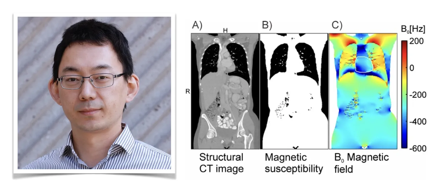
The development of B0 shimming methods relies on the B0 conditions in different human subjects, so it is crucial to understand the root causes of B0 magnetic field distributions and variations within the heart. Previously, researchers suggested that B0 variation in the heart is related to magnetic susceptibility differences between heart tissue and lung cavities, as well as structural changes linked to cardiac motion. For the first time, this study shows highly detailed B0 conditions across cardiac cycle consistent with B0 distributions computed based on subjects’ anatomy from first principles of physics considering motion-induced geometry changes. The concept of B0 magnetic field simulation in the human heart is presented in Figure 2 (Shang Y et al. NMR Biomed. 2022;35(8):e4739. https://pubmed.ncbi.nlm.nih.gov/35393706/).
One of the most remarkable aspects of this study is that the validated simulation can be used as an alternative to in vivo measurements for obtaining the B0 conditions in the human heart by using readily available structural images obtained from multiple imaging modalities. Consequently, we can efficiently investigate B0 conditions at a large sample size without using scanners at a high cost and subject effort, allowing us to develop optimal subject-specific and potential population-based B0 shim methods.
 Coronal slice of a thoracic CT image including heart and neighboring lungs. B) The derived magnetic susceptibility distribution separates tissue and bone (white) from air-filled spaces inside and outside the body (black). C) B0 variations throughout the body including the heart (white outline) are calculated with our novel method in a matter of seconds.")