The MR SCIENCE laboratory pursues translational research in the fields of magnetic resonance imaging (MRI), spectroscopy (MRS) and spectroscopic imaging (MRSI) to advance their clinical potential for the study of neurodegenerative diseases. To this end, MR method developments are combined with state-of-the-art MRI, MRS and MRSI techniques to derive in vivo direct knowledge of the pathobiochemistry underlying clinical conditions such as multiple sclerosis, diabetes or post-traumatic stress disorder (PTSD).
Detection sensitivity and spectral dispersion of MRS/MRSI are improved with the application of ultra-high scanner B0 fields. However, true benefits are only achieved if the concomitant methodological and technical challenges can be overcome. As such, our methodological work at the MRRC's 7 Tesla high field MR scanner sets the stage for in vivo clinical research of the human brain.
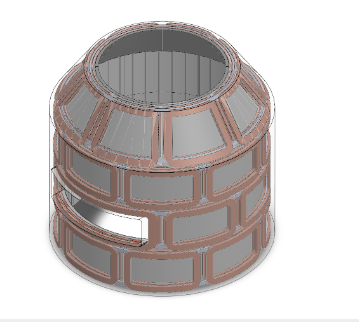
To date, MRI depends on large scanner hardware in which the subject must remain motionless for long periods in a very narrow and enclosed space. This hinders studies on motor behavior and social interactions with other individuals in a natural, upright position. The goal of this research is to develop and construct a Multi-Coil array that enables simultaneous B0 shimming and imaging in the presence of high B0 inhomogeneity in a portable head-only 1.5 T scanner with minimal mobility restrictions.
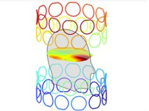
Excellent magnetic field homogeneity is essential for meaningful MRI, MRS and MRSI applications. We have developed a novel method for the correction of magnetic field distortions that is based on the combination of simple, localized coil fields. This multi-coil approach allows to achieve unrivaled levels of field homogeneity in the human brain in a safe way.
The best MR methods are essential for meaningful translational and clinical research. Reliable and stream-lined processing software is necessary for efficient experiment planning and data monitoring during experimental sessions. Finally, robust tools for the analysis of the obtained data are required, e.g. for the quantification of neurochemicals from an in vivo MR spectrum.
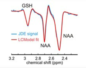
Multiple sclerosis is a chronic disorder of the central nervous system that leads to demyelination and neurodegeneration. Despite enormous research progress, the cause and the underlying neurobiological mechanisms remain poorly understood, thereby preventing improved treatment or the development of a true cure. Magnetic resonance spectroscopy allows to assess the pathological changes and brain damage from the earliest stage.
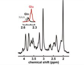
The human brain is a highly dynamic and complex chemical environment in which metabolic homeostasis and brain function rely on the sophisticated interplay of a wealth of neurochemicals. Quantitative information on the involved substances is necessary to study ongoing processes and underlying mechanisms. More importantly, a number of neurological dysfunctions and clinical conditions are reflected by the tissue's neurochemical composition.
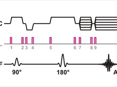
To date, spatial encoding for MRI is based on linear X, Y and Z field gradients generated by dedicated X, Y and Z wire patterns. The multi-coil (MC) technique developed at the Yale MRRC enables the generation of a multitude of magnetic field shapes relevant to biomedical MR. We recently introduced the Dynamic Multi-Coil Technique (DYNAMITE) for MRI and presented proof-of-principle implementations of radial and algebraic DYNAMITE MRI.
