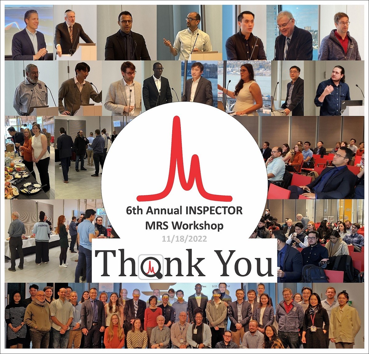
Sixth Annual INSPECTOR MR Spectroscopy Workshop
Columbia University in the City of New York
Friday, November 18, 2022, 8:30am – 5:30pm EST, Jerome L. Greene Science Center (JLGSC) 9th floor
8:30 AM Welcome & Agenda
Christoph Juchem, Ph.D., Columbia University
8:45 AM Fulfilling a pressing need for standardization in MRS research: MRS-BIDS and NIfTI-MRS
Mark Mikkelsen, Ph.D., Weill Cornell Medicine
In vivo MRS is an increasingly utilized biomedical imaging modality for the noninvasive detection of molecules in the human body. However, it lags behind other MR techniques in terms of standardization of methodological approaches. Namely, there is enduring heterogeneity in data acquisition, analysis, reporting, and sharing methods. In this talk, I will present recent initiatives undertaken by the MRS community to move toward standardizing our science. I will briefly introduce the Brain Imaging Data Structure (BIDS), a standard for organizing neuroimaging data, and then speak on extending this standard to include MRS data and metadata, denoted MRS-BIDS. This extension incorporates the new NIfTI-MRS data format, which I will also describe. These initiatives are just two that will aid in the further standardization of MRS research.
9:15 AM Getting to brain mechanisms of injury and disease in vivo: unmet needs
Michael L. Lipton, M.D., Ph.D., Albert Einstein College of Medicine
MRI offers unprecedented access to the living human brain and has facilitated detection of where pathology was previously undetectable in vivo (e.g., demyelinating diseases) or thought to not exist at all (e.g., concussion, excitotoxicity, plasticity). Clinical research has substantiated the import of imaging signatures by relating them to functional outcomes, such as cognitive performance. To ultimately develop effective interventions to address injury and disease requires specific insight into the cellular and molecular mechanisms, such as inflammation, underlying imaging features. This talk will place target mechanisms in context, with suggestions for how MRS could be applied to advance mechanistic discovery.
9:45 AM Improvement of Brain MRS at 7T Using a Wireless Radiofrequency Array
Akbar Alipour, Ph.D., Mount Sinai School of Medicine
Magnetic resonance spectroscopy (MRS) can particularly benefit from substantial enhancement in SNR and spectral resolution at ultra-high field (UHF ≥7T), enabling improved quantification of metabolites. However, at 7T wavelength effects cause a highly inhomogeneous transmit magnetic field in the human brain, with lower transmit efficiency in the posterior-fossa manifesting as signal dropout in this region. Recently, we reported advantages of a surface-applied inductively-coupled radiofrequency array to improve transmit efficiency and signal sensitivity at 7T MRI focusing on cerebellum and inferior temporal lobes. Here we demonstrate the feasibility and effectiveness of in-vivo MRS using the array in human cerebellum at 7T.
10:15 AM Coffee Break
10:45 AM Uncertainty propagation in absolute metabolite quantification for in vivo MRS of the human brain
Ronald Instrella, M.S., Columbia University
Absolute spectral quantification in proton magnetic resonance spectroscopy (1H-MRS) is the standard method for deriving concentration estimates from metabolite signals in absolute units, such as molar concentration. Derived concentrations are often reported with minimum variance estimators, specifically the Cramér-Rao Lower Bound (CRLB), which serves as a proxy for the standard deviation. However, characterizing the precision of absolute quantification relies on more than the CRLB of the metabolite’s signal amplitude from MR signal models. In this study, we present an analysis using the law of propagation of uncertainty to examine this additive effect, and calculate a more comprehensive estimate of the overall precision from concentrations derived using an internal water reference.
11:15 AM Downfield MRSI in the human brain
Peter Barker, Ph.D., Johns Hopkins University School of Medicine
The resonances downfield from water in proton MRS spectra of the human have been under-studied compared to those upfield, in part because they predominantly originate from exchangeable protons that are saturated by conventional water suppression sequences such as VAPOR. However, they may be readily observed using pulse sequences that avoid water saturation, as has been previously demonstrated by several groups using single voxel localization techniques. This presentation will demonstrate feasibility of mapping downfield exchangeable amide resonances in the human brain at both 3T and 7T, and also preliminary data in patients with brain tumors.
11:45 AM Downfield spectroscopy at ultrahigh field at the University of Pennsylvania
Neil Wilson, Ph.D., University of Pennsylvania
Long has in vivo proton spectroscopy focused on the upfield side of water where the majority of the metabolites resonate. This is partially because of the challenges facing spectroscopy in the downfield such as low signal, cross relaxation or chemical exchange with water, and short T2 values. Nevertheless, certain metabolite biomarkers can only be accessed downfield. Here, we present some methods to overcome these technical challenges and highlight applications looking at resonances of NAD+, tryptophan, and carnosine as well as cross relaxation rate measurement of adenosine-containing compounds.
12:15 PM Open Discussion: Moving MRS methodological consensus to acceptance within the clinical research community - A first assessment of dissemination status quo
Moderator: Jodi Weinstein, M.D., Stony Brook University School of Medicine
Moderator: Christoph Juchem, Ph.D., Columbia University
12:45 PM Lunch Break
1:15 PM MRS spectral registration using deep learning
David Ma, M.S., Columbia University
A novel convolutional neural network based spectral registration approach is introduced to perform simultaneous frequency-and-phase correction (FPC) of single-voxel MEGA-PRESS magnetic resonance spectroscopy (MRS) data. For this approach, one neural network was trained and validated using a published simulated and in vivo MEGA-PRESS MRS dataset with a wide range of artificial frequency and phase offsets applied. Our proposed approach was subsequently tested and compared to current sequential FPC deep learning approaches. Our results demonstrate the utility of using deep learning for spectral registration and the application of unsupervised learning in further improving the model to attain state-of-the-art performance.
1:45 PM Lower global N-acetylaspartate associated with illness duration, suggests accelerated aging in persons with psychosis – MRI and whole-brain proton MR spectroscopy at 3 T
Oded Gonen, Ph.D., New York University Langone Medical Center
Persons with schizophrenia and related psychoses (SRP) often show cognitive deficits and early dementia. To test for molecular underpinnings, we compared whole-brain measures: N-acetyl-aspartate (NAA) concentration (WBNAA) - for neuronal health; gray and white matter volume fractions (fGM, fWM) in SRP, nonpsychotic affective patients and healthy controls, to test whether WBNAA will: (i) be lowest in SRP; and (ii) corelate with disease duration (DD). These tests revealed that SRP suffered significant 7% greater GM atrophy than controls (0.43±0.07 versus 0.46±0.04) but the affective cohort did not differ. The fWM, 0.34±0.03, did not differ among all cohorts. DD negatively predicted WBNAA in both SRP and affective cohorts (r= –0.42 and –0.29) suggesting diffuse neuronal dysfunction twice faster than normal aging.
2:15 PM Imaging uptake and metabolism of deuterated choline in rodent models of glioblastoma
Henk De Feyter, Ph.D., Yale University
Choline is a vitamin-like molecule that is almost exclusively obtained through diet. It is an essential component in the synthesis of phospholipids which are key building blocks of cell membranes. In proliferating tissue, such as growing tumors, demand for choline is often increased, resulting in upregulated choline transport and metabolism. Here we explore the use of deuterium metabolic imaging (DMI) to detect the uptake and metabolism of 2H-labeled choline as imaging biomarker of tumor burden with high tumor-to-brain image contrast, following intravenous infusion in rodent models of glioblastoma.
2:45 PM Joint spectral quantification of MR spectroscopic imaging using linear tangent space alignment-based manifold learning
Chao Ma, Ph.D., Harvard Medical School
Spectral quantification is a critical step in quantitative MRS/MRSI. This problem has been extensively investigated in the literature, leading to several widely used methods such as LCModel and QUEST, which take advantage of spectral prior information for quantification. Recent advances in MRSI have made high-resolution MRSI practical in clinical settings. This, in turn, opens new opportunities for imposing both spatial and spectral prior knowledge of MRSI data to improve spectral quantification. This work presents a new method for MRSI spectral quantification using linear tangent space alignment-based manifold learning.
3:15 PM Coffee Break
3:45 PM Clinical translation of hyperpolarized 13C MRS
Kofi Deh, Ph.D., Memorial Sloan Kettering Cancer Center
Several diseases, such as cancer and Alzheimer’s disease, exhibit changes in metabolism which, if quantified, can improve early detection or classification. Current clinical metabolic imaging techniques such as positron emission tomography (PET) and proton magnetic resonance spectroscopy (MRS) have shortcomings of specificity and sensitivity respectively. Hyperpolarized (HP) 13C magnetic resonance spectroscopy (MRS) benefits from a 10000-fold increase in signal to noise ratio compared to thermally polarized MRS and promises to overcome these shortcomings. However, it has unique challenges. We discuss some of these challenges and the innovations being pursued to make HP 13C imaging into a routine diagnostic modality.
4:15 PM Siemens MR spectroscopy
Sinyeob Ahn, Ph.D., Siemens Healthineers
Siemens continue to endeavor to develop MR spectroscopy for research needs and clinical applications. This includes recent developmental trends, including fast imaging acquisition/fast reconstruction, robust data acquisition, and clinical validations. For example, automated easy-to-use single voxel spectroscopy would help reduce scan-to-scan variations while reducing the MRS workflow complexity thus, making the scan operation simpler. Spectroscopic imaging for the glioblastoma application will add its unique clinical values which may not be provided in conventional contrast-based imaging techniques. Reconstruction platform such as the FIRE will help researchers to implement own developments relatively easily, without relying on online reconstruction using ICE.
4:45 PM Unravelling bone marrow adipose tissue composition in proximal femur through 3T MRS
Dimitri Martel, Ph.D., New York University Langone Medical Center
Bone marrow adipose tissue (MAT) is a dynamic endocrine tissue closely involved in bone remodeling. Recently, it has been shown that fatty acids produced by MAT adipocytes can have lipotoxic effects and are associated with increased fragility risk, notably in osteoporosis. The aim of this presentation is to provide an overview of the fatty acid composition of MAT using MRS, its implication on bone quality, and present our recent work on osteoporosis.
5:15 PM Final Comments & Adjournment
Christoph Juchem, Ph.D., Columbia University

