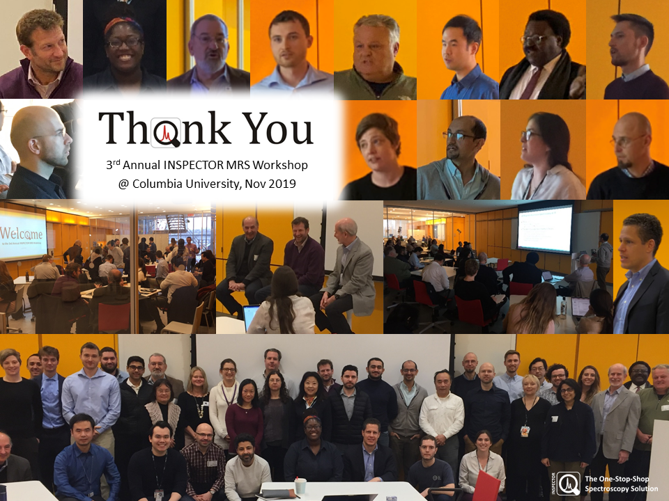
Third Annual INSPECTOR MR Spectroscopy Workshop
Columbia University in the City of New York
Jerome L. Greene Science Center
3227 Broadway (West 129th and Broadway), New York, NY 10027
Friday, 11/22/2019, 8:30 am - 6:00 pm, conference room L3-079 (3rd floor)
Scheduled Program
8:30 AM - 8:45 AM
Welcome and agenda
Christoph Juchem, Ph.D., Columbia University
Brain metabolite mapping using MRSI – Methods and applications
Michal Považan, Ph.D., Johns Hopkins University
Magnetic resonance spectroscopic imaging (MRSI) is a non-invasive technique that merges the principles of magnetic resonance spectroscopy (MRS) and magnetic resonance imaging (MRI). It provides spatially resolved metabolite concentrations from a region of interest. The temporal (i.e. speed) and spatial resolution of an MRSI experiment is always influenced by the available signal-to-noise ratio. This talk covers the current efforts to achieve a high spatial resolution with sufficient SNR in feasible scan time. Advantages of a so-called FID-MRSI method are closer analyzed. Various methods of acceleration such as parallel imaging and spatial-spectral methods are described in detail. We find out why the concentric circles appears to be the best method of acceleration for MRSI (but not MRI). Water and lipid signals and B0/B1 field inhomogeneities may substantially deteriorate MRSI spectral quality. Therefore, the recent methods of water and lipid suppression used in MRSI are presented. Thanks to an increased spectral resolution and higher SNR, MRSI benefits from ultra-high field (³7T). However, novel approaches are necessary to overcome the technical challenges that arise at such a high magnetic field strength. Ultimately, we take a closer look on ultra-high field MRSI methods and some of the advantages of the parallel transmission.
The relationship between hippocampal cerebral blood flow, white matter hyperintensities and
neurometabolites in middle-aged adults
Kay Igwe, M.Sc., Columbia University
The hippocampus is a structure affected by Alzheimer’s disease (AD). Although AD symptoms emerge in later life, increasing evidence suggests that early neurobiological changes associated with AD occur in mid-life. Neuroinflammation has been implicated in the pathogenesis of AD. Neurometabolites such as Choline (Cho), Glutamine and Glutamate (Glx), and Inositol (Ins), may reflect aspects of neuroinflammation and neurodegeneration within the hippocampus. The purpose of this study was to examine the relationship between cerebral blood flow (CBF) and neurometabolite levels in the hippocampus among middle-aged adults and white matter hyperintensity (WMH) volume. We hypothesized that increased markers of neuroinflammation and neurodegeneration would be associated with lower hippocampal CBF and higher WMH volume.
Two-dimensional spectroscopy at ultrahigh field: COSY and JSASSI
Gaurav Verma, Ph.D., Mount Sinai School of Medicine
Even at ultrahigh field, metabolites like gamma-aminobutyric acid (GABA), glutathione (GSH) and glutamate/glutamine compete with overlapping resonances from higher-concentration metabolites within the proton spectrum. Two-dimensional correlated (COSY) and J-resolved (JPRESS) spectroscopy introduce a second spectral dimension to resolve these peaks. Together with sensitivity and spectral separation advantages using ultrahigh field MRI, these techniques can resolve metabolites difficult to reliably quantify using conventional techniques and lower field strengths.
7T COSY employs a 90-180-Δt1-90 pulse sequence to generate a coherence-transfer echo with an incrementing time delay (Δt1). Following double Fourier transform, COSY spectra resolve into directly-sampled (F2) and indirectly-sampled (F1) spectral dimensions. Non-coupled metabolite peaks appear along a main diagonal trend where F2=F1, but J-coupled peaks experiencing coherence transfer during t1 evolution appear as “cross peaks” with position dependent on the chemical shift of both coupled spins. The COSY technique was implemented to resolve 2-hydroxyglutarate (2HG) an “oncometabolite” indicative of IDH-mutation in brain tumors.
J-resolved semi-adiabatic spectral-spatial imaging (JSASSI) uses a 90-180-Δt1/2-180-Δt1/2 pulse sequence, and incorporates adiabatic refocusing pulses to mitigate B1-inhomogeneity. JSASSI resolves metabolites according to chemical shifts in the F2 dimension and coupling constants in F1. Implementing adiabatic pulses and a second refocusing pulse implies a better-than-double SNR improvement versus COSY, and may provide reliable quantification of low-concentration metabolites otherwise subject to spectral overlap, such as GABA.
Overview of INSPECTOR software
Christoph Juchem, Ph.D., Columbia University
Magnetic resonance spectroscopy (MRS) is a powerful tool for clinical research and clinical diagnostics of virtually all disorders that possess distinct metabolic signatures. The pursuit of optimal experimental quality should be complemented by similarly optimal data processing and quantification strategies and the relevance of advanced yet user-friendly analysis software for the reliable extraction of biochemical information cannot be overstated. The recently introduced software INSPECTOR comprises comprehensive processing and analysis functionality for in vivo MRS in a one-stop-shop solution with data handling, quality management, linear combination modeling and visualization capabilities. I here present INSPECTOR as tool for biomedical research and discuss its suitability for interactive and hands-on learning of MRS processing and quantification strategies. INSPECTOR provides maximal transparency at every step in the MRS processing chain along with ample documentation, protocol capabilities, export options and a user-friendly interface. INSPECTOR is available to academic institutions free of charge.
Optimizing MEGAPRESS processing in translational magnetic resonance spectroscopy study in
rodent models
Jia Guo, Ph.D., Columbia University
1H MRS spectral editing of GABA with MEGAPRESS is seeing increasing popularity in human studies thanks to the recent implementation of standard pulse sequences and processing algorithms. Nevertheless, in preclinical studies, rodent models continue to play a significant role in the scientific investigation of neuropsychiatric disorders. However, unlike for human studies, there has been a lack of similar standardization for small animal studies. With the continued development of transgenic rodent models of human brain disease, there is an increasing need to measure and analyze GABA‐edited MR spectra in rodents routinely. In this talk, I will present an automated MEGAPRESS processing pipeline tailored for in vivo rodents studies that is designed to enable an unbiased evaluation of MR spectra through Spectrum Registration. A review of in vivo MEGAPRESS study in rodent models will also be covered.
To Twix or not to Twix: Processing of Siemens MEGA-PRESS data made easy
Dikoma Shungu, Ph.D., Cornell University
Siemens MEGA-PRESS data exported in so-called ‘rda’ format are simple to process because the scanner automates the combining of multi-channel and separation of edit-on and edit-off data into two single regular FID signals that can be filtered, Fourier-transformed and subtracted to obtain the desired J-edited spectrum. However, there is no option to examine and correct the individual averages for Bo or phase shifts, often leading to poor-quality edited spectra. By contrast, while processing of multi-channel MEGA-PRESS data exported with the ‘twix’ utility enables individual averages to be corrected for Bo and phase shifts, the raw signals from individual phased-array coil elements must first be combined before they can be corrected for Bo and phase shifts, separated into the edit-on and edit-off subspectra and then subtracted to obtain the final edited spectrum, which can be computationally quite complex.
For this presentation MEGA-PRESS data exported in standard DICOM format will be processed interactively and demonstrated to offer an effective compromise between the limitations of rda-exported and complexity of twix-exported Siemens MEGA-PRESS data.
Panel Discussion: What do we want in a MRS software?
Panelist: Lawrence Kegeles, M.D., Ph.D., Columbia University / New York State Psychiatric Institute
Panelist: Douglas Befroy, D.Phil., PeakAnalysts Inc.
Panelist: Douglas Rothman, Ph.D. Yale University
Moderator: Jodi Weinstein, M.D., Stony Brook University
A functional magnetic resonance spectroscopy study using chemogenetics
David Guilfoyle, Ph.D., Nathan Kline Institute
In this study we use “designer receptor exclusively activated by designer drugs” (DREADDs) chemogenetic technology for site-specific neural activation in the rat pre-frontal cortex at 7T. Single voxel spectroscopy was used to measure the changes in the neurochemical profile caused by the chemogenetic stimulation. We also measured changes in cerebral blood flow (CBF). The strongest and most significant metabolite concentration changes were in N-acetylaspartate (NAA) and lactate.
MRS of liver metabolism
Martin Gajdošík, Ph.D., Columbia University
The liver is the largest gland and, after skin, the second largest organ in the human body. The hepatic cells contain glycogen, fat, and iron compounds and their external secretion is bile. 1H MR spectroscopy is currently one of the most precise non-invasive methods to quantify hepatic fat and it is also feasible to use this method to detect iron and glycogen. In combination with 13C and 31P MR spectroscopy, this method can deliver even more detailed information about liver metabolism. Overview of the methods and current work on liver MR spectroscopy at MR Science Laboratory will be presented.
Hyperpolarized 13C metabolic imaging: Basic principles and applications
Dirk Mayer, Ph.D., University of Maryland
Dissolution dynamic nuclear polarization (dDNP) has been developed to overcome the intrinsic low sensitivity of MR-based metabolic imaging. This presentation describes the basic principles of dDNP, the special imaging considerations due to the nonrecoverable nature of the increased magnetization, and various in vivo applications including cancer, traumatic brain injury, and liver disease.
Tales from the flip side: A non-traditional career in MR research
Douglas Befroy, D.Phil., PeakAnalysts Inc.
The development of novel MR methods to investigate metabolism in vivo is accompanied by challenges – both physiological and MR related – that require innovative solutions. My research has encompassed a variety of paradigms: from plants to humans, 1H and X-nucleus spectroscopy, and in vitro to in vivo applications, and this unorthodox background has proved indispensable in my career as an academic and as an MR-consultant who is hired to solve technical problems and resolve methodological issues. In this talk, I’ll discuss some of the challenges and solutions that I’ve encountered along the way.
Detecting Glx and GABA in the hippocampus at 3T using semi-LASER MRS: Feasibility in patients
with schizophrenia
Jodi Weinstein, M.D., Stony Brook University
In effort to examine excitation/inhibition balance in the hippocampus of patients with schizophrenia, 1H‑MRS at 3T was used to measure the combination of glutamate and glutamine (Glx) and GABA neurometabolites. The hippocampus has traditionally been a challenging region to measure with 1H‑MRS because of its relatively small size and its location deep in the brain. Several 1H-MRS protocols were tested to maximize signal-to-noise ratio while maintaining regional specificity of hippocampal tissue. Feasibility results for the use of (MEGA-)sLASER sequence will be presented.
Advanced 3D MRSI in the hippocampus in patients with schizophrenia
Oded Gonen, Ph.D., New York University
Hippocampal disruption may be a central pathology for psychosis as many of its measures differ between schizophrenia cases and healthy controls, e.g., reduced volume, increased resting blood flow, impaired task-related activation, decreased neurogenesis and reduced connectivity with other regions. As the disease entails cognitive and attention deficits, effort-independent methods, e.g., MR modalities, especially 3-dimensional proton MR spectroscopic imaging (3D 1H-MRSI), are well suited for its investigations. 1H-MRSI noninvasively yields metabolic markers of several cellular processes, most notably: NAA (N-Acetylaspartate and N-acetylaspartylglutamate) for neuronal integrity; Cr (phosphocreatine and creatine) for energy metabolism; Cho (choline, phosphocholine and glycerophosphocholine) for membrane turnover and astroglia proliferation; and myo-inositol (mIns) for inflammation and gliosis. Using 3D 1H-MRSI to cover the entire bilateral hippocampi, we show patients’ average hippocampal Cr concentration was 19% higher than controls’, 8.7±2.2 versus 7.4±1.2 mM (p‹0.05); with no differences in NAA: 8.8±1.6 vs. 8.7±1.2 mM, Cho: 2.33±0.7 vs. 2.1±0.3 mM, or mIns: 6.12±1.46 vs. 5.19±0.88 (all p›0.1). There was a positive correlation between mIns and Cr in patients (r=0.57, p=0.05) but not controls. Bilateral hippocampal volume was ~10% lower in patients: 7.5±0.9 vs. 8.4±0.7 cm3 (p‹0.05). These findings suggest that the hippocampal volume deficit in schizophrenia is not due to net loss of neurons, in agreement with histopathology studies but not with prior 1H-MRS reports. Elevated Cr is consistent with hippocampal hypermetabolism and its correlation with mIns may also suggest an inflammatory process affecting some cases; this may suggest treatment targets and markers to monitor them.
Evidence-based 1H MRS spectral fitting and quantification: Application to multiple sclerosis
Kelley Swanberg, M.Sc., Columbia University
1H MRS is a method unparalleled in its promise to noninvasively profile the neurochemistry behind many neurological disorders, among them the autoimmune demyelinating condition multiple sclerosis. Despite the method's potential, however, the research and especially clinical utility of 1H MRS data sets depends heavily on their influence by real biological phenomena as opposed to experimental error, common sources of which include suboptimal spectral processing, fitting, and quantification procedures. In this talk I will introduce the latest research and updates to INSPECTOR that enable evidence-based 1H MRS data fitting and quantification, especially as pertains to the past and future of 1H MRS investigations into multiple sclerosis progression and disease-modifying therapies.
Realistic and efficient simulation of spectral basis sets
Karl Landheer, Ph.D., Columbia University
Accurate quantification of magnetic resonance spectroscopic experiments requires the simulation of basis sets which are linebroadened and scaled via linear combination modelling. It is best practice to use physical parameters (RF/gradient waveforms, timings) that are physically representative of experimental reality, as well as a sufficient number of spatial points across the voxel and voxel boundaries to accurately reflect the continuous reality. A novel software packaged, referred to as Magnetic Resonance Spectrum Simulator (MARSS), will be discussed which can be used to simulate a full basis set in ~30 minutes on a desktop computer. A brief overview of the relevant physics will be discussed, which includes the quantum mechanical density operator and coherence order transfer pathways. Examples of how to use MARSS for a custom pulse sequence will be discussed, as well as the resulting high quality fits obtained with INSPECTOR will be shown across a variety of pulse sequences and field strengths.

