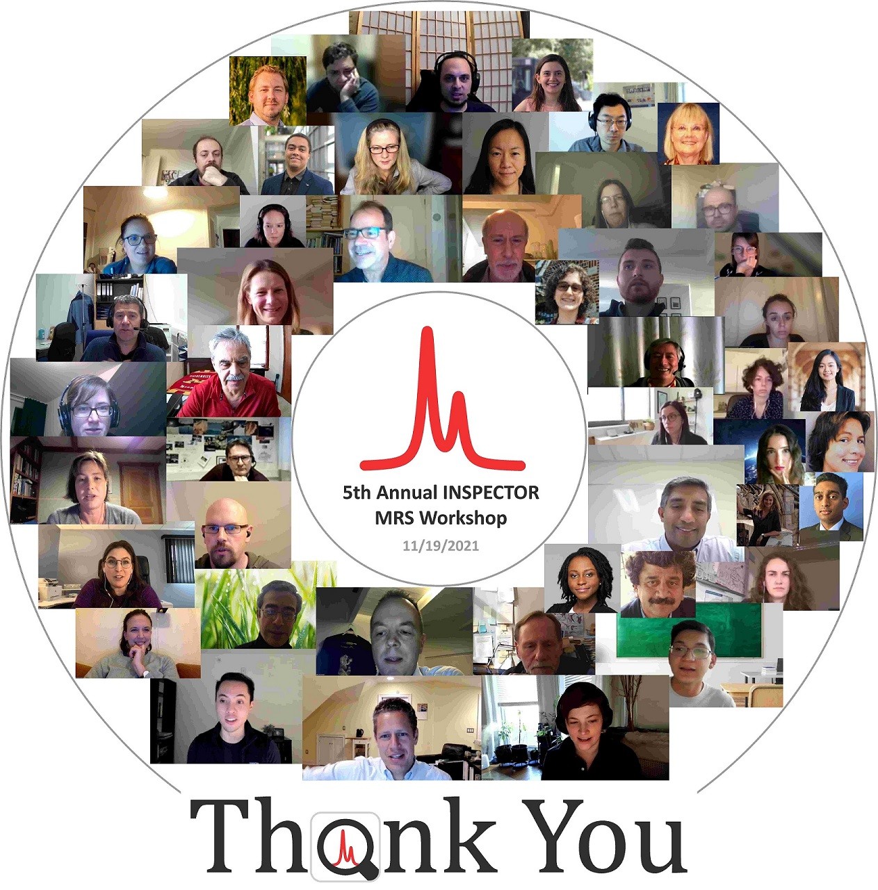
The MR SCIENCE Laboratory presents the 5th Annual INSPECTOR Magnetic Resonance Spectroscopy Workshop at Columbia University in the City of New York.
Fifth Annual INSPECTOR MR Spectroscopy Workshop
Columbia University in the City of New York
Friday, 11/19/2021, 8:30 am - 5:30 pm, Virtual meeting
8:30 AM
Welcome and agenda
Christoph Juchem, Ph.D., Columbia University
8:45 AM
Macromolecules and baseline in short and intermediate TE proton MR brain spectra
Cristina Cudalbu, Ph.D., École Polytechnique Fédérale de Lausanne (EPFL)
Proton MR spectra of the brain measured at short and intermediate echo times (TE) contain signals from mobile macromolecules (MM). In normal brain, MM signals arise mainly from the protons of amino acids within cytosolic proteins. These broad peaks of MM underlie the narrower peaks of metabolites and often complicate their quantification but they also may have potential importance as biomarkers in specific diseases. Thus, reliable detection and fitting of MM are crucial for accurate quantification of brain 1H-MR spectra. Higher spectral resolution at ultra-high field (UHF) led to attempts to understand the origin of the MM spectrum, to decompose it into individual spectral regions or peaks and to use the components of the MM spectrum as markers of various physiological or pathological conditions. The aim of this presentation is to provide an overview and some recommendations on how to handle the MM signals and the baseline in different types of studies.
9:15 PM
Decreased GABA and glutamate concentrations in the visual cortex via 1H MRS at 3 T and reduced fMRI activation in
the fusiform face area induced by pharmacologically increased dopamine levels
Ralf Mekle, Ph.D., Center for Stroke Research Berlin, Charité Universitätsmedizin Berlin
Schizophrenia frequently manifests psychotic symptoms, such as delusions and hallucinations. However, its neurochemical mechanisms are not well deciphered, though dysfunctional dopaminergic neurotransmission is suggested. In this study, single volume 1H MRS using MEGA-PRESS at 3 T was applied to investigate neurochemical changes in the visual cortex induced by pharmacologically increased dopamine levels in healthy volunteers that also performed a visual detection task monitored by fMRI. Increased dopamine yielded decreased GABA and reduced glutamate, as well as diminished fMRI activation in the fusiform face area. Results support the theory that glutamate hypofunction might contribute to the formation of hallucinations in schizophrenia.
9:45 AM
Muscle MRS in muscular dystrophy.
Hermien Kan, Ph.D., Leiden University Medical Center
Muscular dystrophies are characterized by progressive muscle weakness. Clinically, this results in functional decline and loss of muscle function. On the tissue level, muscles are replaced by fat and fibrotic tissue, representing end stage disease, and metabolic alterations and inflammation, which are present earlier in the disease process. While fat and fibrotic changes are considered irreversible, measures of inflammation and especially metabolism reflect muscle tissue that still functions and is therefore potentially amenable to treatment. In this talk, I will present an overview of MR spectroscopy work that has been done in muscular dystrophies, and highlight possible future directions.
10:15 AM
Coffee break
10:45 AM
1H Magnetic Resonance Spectroscopy in the assessment of brain tumors
Eva-Maria Ratai, Ph.D., Harvard Medical School
The evaluation of brain tumors is one of the areas where 1H MRS has impacted patient management significantly. Glioblastomas are challenging cancers to treat, and long-term favorable clinical outcomes in patients with recurrent glioblastoma (rGBM) continue to be difficult to achieve. One of the main characteristics of glioblastoma is the presence of intratumoral neoangiogenesis. Therefore, patients with rGBM are often treated with anti-angiogenic agents such as bevacizumab. Because the use of anti-angiogenetic therapy is frequently associated with substantial reduction in contrast enhancement on T1-weighted MR imaging, it is often difficult to distinguish between a true favorable tumoral response from so called pseudo-response using conventional MRI. Our data suggests that MRS may be a useful modality to predict the efficacy of anti-angiogenic therapy to distinguish true tumor response from pseudo-response.
11:15 AM
Diffusion Weighted Magnetic Resonance Spectroscopy (DW-MRS): Cell physiology meets microstructure
Itamar Ronen, Ph.D., Leiden University Medical Center
Sensitization of magnetic resonance spectroscopy (MRS) sequences to diffusion with the addition of diffusion-weighting magnetic field gradients lets intracellular metabolites become probes for intracellular morphology. The diffusion properties of metabolites measured with a variety of diffusion weighting schemes thus provide a unique, cell-type specific microstructural information on the intracellular milieu in a wide range of spatial scales, ranging from cytoplasmic properties to cell morphological features. In this brief presentation, I will give examples of insights obtained from diffusion weighted MRS experiments on neural cell morphology, and provide evidence for the sensitivity of DW-MRS to alterations in cell morphology linked to specific pathomechanisms.
11:45 PM
Integrated data processing pipelines - Compass framework
Jedrek Burakiewicz, Ph.D., Tesla Dynamic Coils
MRS and CSI data processing on commercially available scanners is often very basic and custom processing steps tend to be necessary. This often forces the user to build a pipeline from zero themselves, or to integrate various external processing tools, which can be written in different programming languages and use different data formats. To solve this problem we present Compass, a data processing framework which allows the user to build pipelines that combine built in processing steps, external tools and custom code, and to make it easy to run them with the use of a simple GUI.
12:15 PM
Panel Discussion:
There is no ‘i’ in ‘spectroscopy’: How should we distinguish our research in an increasingly collaborative world?
Panelist: Dennis Klomp, Ph.D., UMC Utrecht
Panelist: Jodi Weinstein, M.D., Stony Brook School of Medicine
Panelist: Karin Markenroth Bloch, Ph.D., Lund University
Panelist: Uzay Emir, Ph.D., Purdue University
Moderator: Kelley Swanberg, M.Sc., Columbia University
In this age of international multicenter clinical trials and genomics databases that cover entire countries, in vivo MR spectroscopy researchers have begun to jump on the bandwagon of large-scale collaborations, from field-wide consensus publications and multi-center analyses to multi-vendor sequences and code and data sharing study-group committees. Against this growing scale of multi-actor endeavors stretching over a framework of funding support still largely awarded at most to a few individual researchers or research centers, what is the extent to which it is feasible, functional, and ultimately desirable to supplement or even replace competitive differentiation with sharing, openness, and exchange? Are the days of the lone spectroscopist hacking away at their own personalized toolkits truly numbered? Or is it unrealistic to expect that the individual researchers with the widest networks of unreserved collaboration will not be left unrecognized and unsupported by the sources of funding and institutional support on which we all still depend? More broadly, what does our ability as a field to promote collaboration instead of competition mean for the future of spectroscopy’s impact on other spheres of biomedical research and clinical application?
12:45 PM
Lunch
1:15 PM
Current applications of high performance 7 T MRSI at the Vienna HFMRC
Gilbert Hangel, Ph.D., Medical University of Vienna
Since the development of a 7 T 3D MRSI method that achieves 2-4 mm resolution in 10-20 min scan time, our research group has tested it for multiple applications. This talk will give an overview of our current progress in scans of healthy subjects and patients with neoplasms, refractory epilepsy and multiple sclerosis.
1:45 PM
Accessibility: Making in vivo MR Spectroscopy approachable for newcomers and experts alike through INSPECTOR
Leonardo Campos, Columbia University
Ranging from signal processing and data science to physics, biology, and chemistry, the interdisciplinary nature of MR spectroscopy makes it an enticing and versatile tool that has increasingly attracted scientists and clinicians alike. However, this large swath of necessary background knowledge also provides a barrier of entry. Physical concepts and data processing techniques are thereby presented through INSPECTOR in an easily interpretable way in order to facilitate accessibility for newcomers to the field.
2:15 PM
Long-term behavioral effects of ultra-high magnetic fields
Ivan Tkác, Ph.D., University of Minnesota
Chronic exposure to static, ultra-high magnetic fields might not be as innocent as we believed. Long-term behavioral changes, especially the tight circling in Morris water maze, were observed in mice chronically exposed to 16.4 T days or even weeks after the last exposure. These finding suggest that the chronic exposure to a magnetic field as high as 16.4 T may result in long-term impairment of the vestibular system.
2:45 PM
Interpreting the in vivo 1H MRS GABA signal as a biomarker of inhibition
Kevin Behar, Ph.D., Yale University
Altered GABA level as detected by 1H MRS is seen in numerous brain disorders and a strong relationship is seen between GABA level, cortical excitability and interconnectivity assessed by EEG and fMRI. While the GABA 1H MRS measurement has gained increasing importance as a biomarker in both clinical and basic neuroscience research, fundamental questions about its meaning remain: Does the GABA MRS measurement provide an accurate assessment of total brain GABA? Do changes in [GABA]MRS reflect similarly in extracellular fluid where binding to GABA receptors occur, and the inhibitory actions are conveyed? In this work we measured brain levels of GABA in rats in vivo using J-edited 1H MRS, in extracts in vitro and in extracellular fluid by microdialysis and mass spectrometry and compared the corresponding change in GABA level from baseline over a range of GABA concentrations using the GABA-elevating drug, vigabatrin. Our findings provide validation of [GABA]MRS measurement and further inform its use as a translational biomarker of inhibition.
3:15 PM
Coffee break
3:45 PM
Are Cramér-Rao lower bounds an accurate estimate for standard deviations in in vivo magnetic resonance spectroscopy?
Karl Landheer, Ph.D., Regeneron Pharmaceuticals
Cramér-Rao lower bounds (CRLBs) have become the stand-in for standard deviations (SDs) throughout the MRS community, however the calculation of CRLBs hinges on several assumptions which are frequently overlooked. In this talk we will systematically investigate these assumptions via realistic simulations where ground truth is known. It was found that CRLBs are an adequate estimator for standard deviation when the model well describes the data. In the case when the macromolecule basis deviates from the measured macromolecules it was shown that the CRLBs can deviate from standard deviations by 50% or more for most metabolites. The implication of these results will be discussed.
4:15 PM
Major Depressive Disorder: Perspectives from proton MRS and ketamine
Larry Kegeles, M.D., Ph.D., Columbia University
The dissociative anesthetic ketamine has rapid therapeutic effect in major depression, and it received FDA approval two years ago as an antidepressant medication. Previously approved antidepressants generally act on monoamine systems – serotonin, dopamine, and norepinephrine – while the major mechanism of action of ketamine is noncompetitive NMDA-type glutamate receptor antagonism. Its novel mechanism has stimulated extensive clinical and preclinical research, and its glutamate system involvement has stimulated investigation with MRS. This talk will present recent work from the Columbia group using proton MRS with J-difference editing to relate acute brain GABA and glutamate changes to clinical effects of ketamine administration.
4:45 PM
MRI of [2-13C] Lactate without J-coupling artifacts
Keshav Datta, Ph.D., Stanford University
Metabolic imaging using hyperpolarized [2-13C]Pyruvate has the potential to simultaneously probe glycolysis and Kreb’s cycle, but one major limitation is the difficulty in imaging [2-13C]Lactate. The peak- splitting induced by the J-coupling between the C2 carbon and its attached proton causes ghosting and blurring artifacts, depending on the k-space trajectory. In this talk, two techniques, the first a two-shot approach combining in-phase and quadrature images acquired at echo times differing by 1/2J and the second a single-shot method employing a highly narrowband radiofrequency excitation pulse that images a single peak from the doublet, are proposed to resolve the J-modulated artifacts.
5:15 PM
Final comments & adjournment
Christoph Juchem, Ph.D., Columbia University
File Upload

