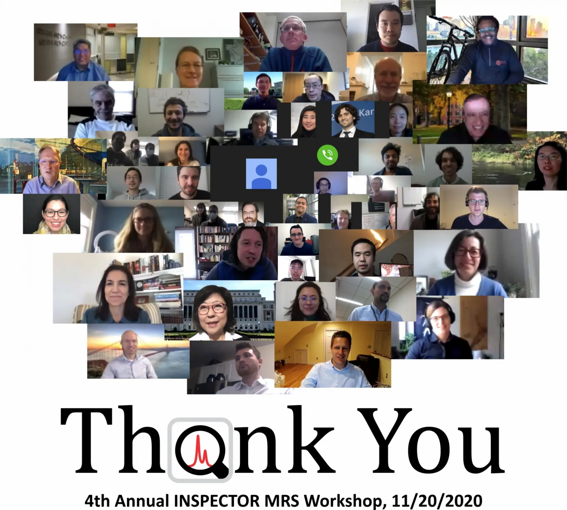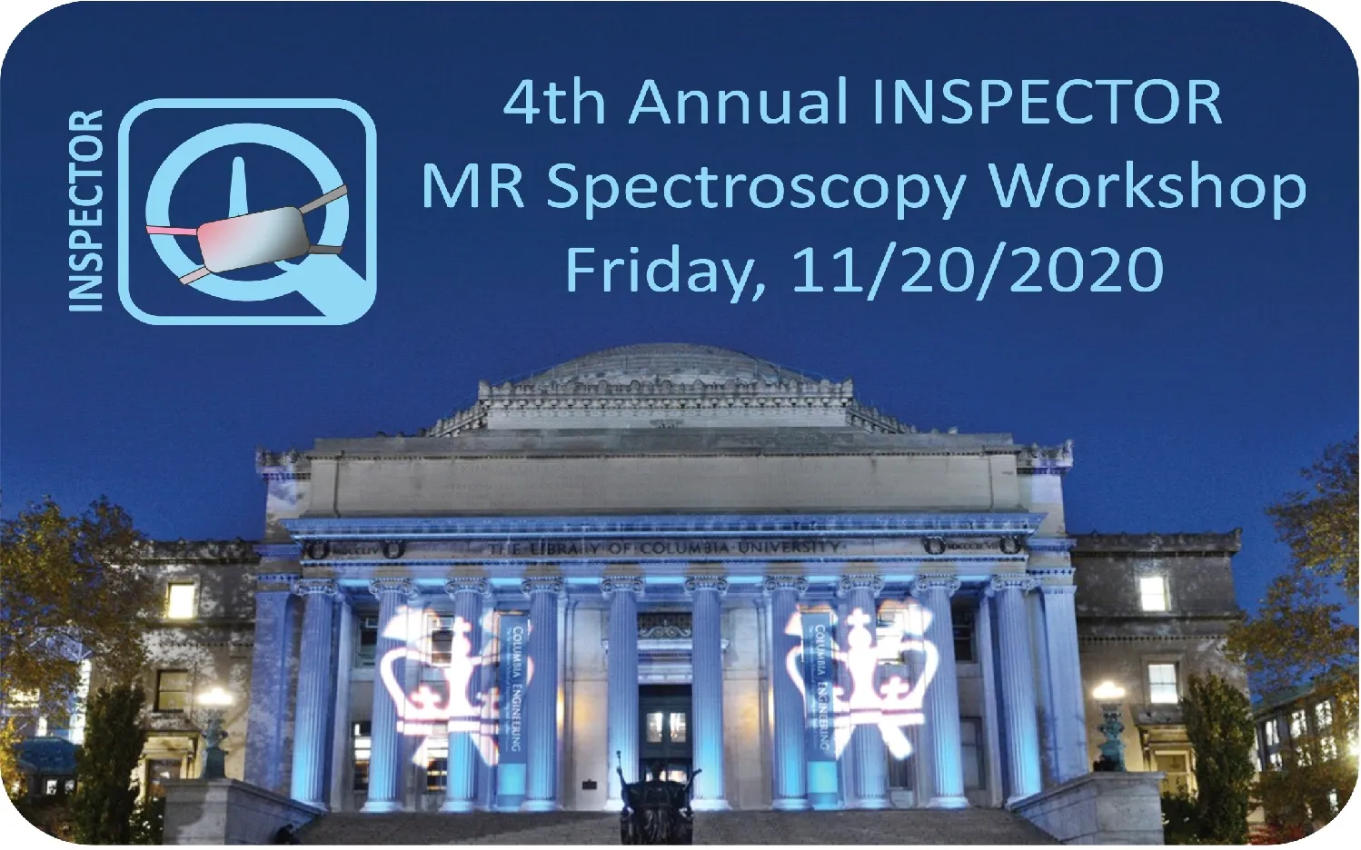
The MR SCIENCE Laboratory presents the 4th Annual INSPECTOR Magnetic Resonance Spectroscopy Workshop at Columbia University in the City of New York. Download the schedule here.
Fourth Annual INSPECTOR MR Spectroscopy Workshop
Columbia University in the City of New York
Friday, 11/20/2020, 8:30 am - 5:20 pm, Virtual meeting
8:30 AM
Welcome and agenda
Christoph Juchem, Ph.D., Columbia University
8:45 AM
Multimodality imaging approach for characterization of pancreatic tumor in a mouse model
Yanping Sun, Ph.D., Columbia University
Pancreatic ductal adenocarcinoma (PDA) is a highly lethal malignancy characterized by oncogenic K-ras and p53 mutations. In this primary study, we used three-dimensional ultrasonography to monitor tumor growth biweekly in 6 PDA mouse models. After the tumors reached 4-7mm diameters, MRI and MRS were performed for characterization of tumor structure and metabolite profiles. Pancreatic tumors showed hypoechoic roughly spherical regions with well-defined border between the PDA and non-PDA tissues. Tumor masses appeared more rigid and retain their overall shape better than normal pancreas by reducing the pressure of the ultrasound transducer on the animal. The tumor showed heterogeneous hyper-intense with small patchy necrosis on T2 weighted MRI. MRS data analyzed with INSPECTOR software depicted lipid metabolites in the tumor with peak assignments (ppm) at 0.9, 1.3, 1.6, 2.02, 2.24, 2.75 and 5.29. Our study demonstrated that Ultrasound, MRI and MRS can be used to characterize the special features of the pancreatic tumor. Evaluating tumor characteristics can help in understanding the PDA biology and in determining efficient treatment approaches.
9:15 PM
7 T MRS of human aging in the default mode network: risk and resilience to Alzheimer's disease
Melissa Terpstra, Ph.D., University of Minnesota
Over the past several years, we have used ultra-short echo time 7 T MRS to measure the neurochemical profile of healthy older adults and participants with Alzheimer’s disease (AD). We used single voxel MRS of the posterior cingulate cortex, i.e., the hub of the default mode network. Whereas published studies focused on MRS of aging and AD, we are currently adding connectome style imaging to this data set. In cooperation with the human connectome project, we are also collecting a wealth of health and behavioral data for these participants. At this stage, we are using MRS from the published aging and AD cohorts to train predictors and inform on the typically aging HCP-A cohort. We have also begun to correlate MRS outcomes with health and behavioral data. In this talk I will focus on key preliminary findings and MRS fitting considerations, as other fundamental MR parameters for studies on aging and AD.
9:45 AM
Investigating medial prefrontal GABA concentrations in alcohol use disorder with MR spectroscopy
Jonathan Wai, M.D., New York State Psychiatric Institute
Chronic and binge alcohol consumption has been found to decrease prefrontal γ-aminobutyric acid (GABA) when measured soon after the initiation of abstinence. The GABAergic system in alcohol use disorder (AUD) has been of increasing interest because of the potential therapeutic effects of medications that act to increase GABA activity. This presentation will review the literature using 1H-MRS to investigate brain GABA in AUD. Preliminary data from our study measuring GABA concentrations in abstinent individuals with AUD undergoing a transcranial magnetic stimulation study will also be presented.
10:15 AM
Coffee break
• Meet and greet
• INSPECTOR clinic
10:45 AM
Accelerating edited MRS
Richard Edden, Ph.D., Johns Hopkins University
This talk will build from the basics of J-difference editing to discuss how multi-metabolite and multi-voxel editing are possible with the pulse sequences HERMES and MEGA-PRIAM. Building upon HERMES, we will discuss the method HERCULES, which relies on a combination of Hadamard-encoded editing and multi-spectrum linear-combination modeling to quantify multiple edited metabolites in a single scan at 3T.
11:15 AM
Cracking into clinical practice: The ENIGMA effort for MR spectroscopy
Alexander Lin, Ph.D., Harvard Medical School
Despite decades of publications, magnetic resonance spectroscopy is not utilized as a conventional diagnostic tool in radiology. This talk will focus on how MR spectroscopy can be used in clinical practice and discuss the barriers that remain for its full adoption by clinicians. We will discuss how current consensus efforts such as the ENGIMA spectroscopy workgroup and ISMRM study group are working towards addressing those issues.
11:45 PM
Experts’ consensus recommendations on advanced MRS techniques: Overview
In-Young Choi, Ph.D., University of Kansas
Recently MRS experts set out to improve the quality of advanced MRS and to promote its adequate uses in a global setting. The major goal was to offer reference guidelines of major advanced MRS techniques through MRS experts’ consensus recommendations. Our collaborative efforts laid out thirteen topics of the current state of the art MRS, including advanced single voxel MRS, neuro MRSI, editing, shimming, water and lipid suppression, motion correction, macromolecules and pre-clinical MRS in neuro MRS, and 31P MRS of skeletal muscle. Our MRS experts and the community also reached consensus on MRS terminology and minimum reporting standards.
12:15 PM
Panel Discussion:
We have consensus, but what do we need for impact?
Panelist: Anissa Abi-Dargham, M.D., Stony Brook University
Panelist: Robert Fulbright, M.D., Yale University
Panelist: Gülin Öz, Ph.D., University of Minnesota
Moderator: Dikoma Shungu, Ph.D., Cornell University
Despite its promise in principle as a source of noninvasively measured biomarkers for research and clinical use, in vivo magnetic resonance spectroscopy continues to arguably exhibit shortcomings in the standardization of its data acquisition, processing, and reporting methodology that could stand in the way of realizing this potential. The last several months have seen the publication of numerous consensus recommendations toward the more standardized implementation of some of these methods. Nevertheless, the effect of these efforts on the quality of in vivo spectroscopy experimentation and its ultimate impact on questions in basic or translational science and especially on clinical practice remains to be seen. We have consensus papers, but do we have consensus? And even if we now have consensus, is this truly enough for impact?
12:45 PM
Lunch
1:15 PM
MR research at Columbia University
J. Thomas Vaughan, Ph.D., Columbia University
1:45 PM
INSPECTOR software - Introduction and overview
Martin Gajdošík, Ph.D., Columbia University
The free software INSPECTOR comprises comprehensive processing and analysis functionality for in vivo MRS in a one-stop-shop solution with data handling, quality management, basis set simulations, linear combination modeling and visualization capabilities. INSPECTOR provides maximal transparency at every step in the MRS processing chain along with ample documentation, protocol capabilities, export options and a user-friendly interface. Ease of use, compatibility with other data formats and data quality control from loading the data to export of analytical results make INSPECTOR a software of choice for wide use of the MRS methods in preclinical and clinical research. This talk will highlight INSPECTOR’s key features most used in MRS research.
2:15 PM
Automated basis set simulation for in vivo MR spectroscopy
Karl Landheer, Ph.D., Columbia University
MARSS is a novel basis set simulation software which uses the density matrix formalism to simulate accurate spectral shapes for quantification or testing novel sequences. This talk will explain common pitfalls when simulating basis sets and briefly outline the theory behind MARSS. MARSS can be used via a standalone executable and has recently been incorporated into INSPECTOR. How to use MARSS via INSPECTOR’s GUI will be covered in detail, which enables the easy simulation of basis sets for common stock sequences.
2:45 PM
Validating linear combination models for 1H MRS quantification
Kelley M. Swanberg, M.Sc., Columbia University
Despite its promise in principle as a means to noninvasively measure in vivo concentrations of biologically relevant small molecules, 1H MRS continues to fall short as a practically useful sensitive and specific clinical tool. In this talk I will overview several features of spectral data analysis both in- and outside INSPECTOR that facilitate the validation of various linear combination modeling approaches to spectral quantification as well as the multivariate statistical interpretation of resultant metabolite concentrations, with an eye to maximizing the sensitivity and specificity of 1H MRS to biologically salient metabolic signatures and thereby the utility of this “experimental” technique to both biomedical research and clinical practice.
3:15 PM
Coffee break
• Meet and greet
• INSPECTOR clinic
Fast Pitch Sessions
3:45 PM
MRSHub – Online Resources for Code & Data Sharing
Georg Oeltzschner, Ph.D., Johns Hopkins University
Advancing the field of MRS can benefit from collaboration, and methodological consensus across research groups. To foster exchange of expertise, methods, and data, the Committee for MRS Code and Data Sharing of the ISMRM MRS Study Group has created the MRSHub. This free online resource for MRS researchers features a central repository to share and find data analysis code, de-identified datasets for testing and benchmarking methods, and a forum covering a wide variety of MRS-related topics.
3:50 PM
In vivo detection of glutathione in the mouse brain at 9.4T using MEGA-PRESS
Jia Guo, Ph.D., Columbia University
MEGA-PRESS sequence has been implemented at 9.4T to detect the glutathione (GSH) levels in the mouse brain. Our preliminary results demonstrate that MEGA‐PRESS is a feasible technique to measure in vivo GSH levels in the mouse brain at 9.4 T, and has good sensitivity to detect regional differences in GSH levels.
3:55 PM
High resolution 7T 3D-MRSI applied to the research of brain pathologies
Gilbert Hangel, Ph.D., Medical University of Vienna
Fast MRSI techniques make it possible to resolve metabolic changes in brain pathologies in greater detail than ever before. We present our current results for tumors, eplilepsy and multiple sclerosis.
4:00 PM
Calibration-free B1 shimming for single-voxel MRS
Adam Berrington, Ph.D., University of Nottingham
Sufficient control of the RF transmit field (B1+) in small regions-of-interest is critical for single voxel MR spectroscopy at ultra-high field. Static RF shimming can improve local B1+, but must be optimised for each participant and ROI, which limits its applicability in practice. By calculating B1+ shim solutions offline over a database of participants, we present a method to obtain shim solutions which can be applied to any participant, to produce a desired |B1+| within pre-defined target ROIs.
4:05 PM
Optimization of spatial-spectral pulses with the adiabatic SLR algorithm
Gaurav Verma, Ph.D., Mount Sinai School of Medicine
The adiabatic SLR algorithm allows optimization of spatial-spectral adiabatic RF pulses to multiple constraints including time, bandwidth, SNR, SAR, and hardware limits. The algorithm was used to design pulses for applications including 2D spectroscopy, GABA-edited spectroscopy and EPSI.
4:15 PM
Semi-LASER implementation on the GE platform
Ralph Noeske, Ph.D., GE Healthcare
The MRS community worked in recent years on finding consensus on techniques that should be used for clinical proton MRS in the brain. For single voxel the recommendation was to replace the currently most widely used PRESS technique by semi-LASER, mainly due to the much lower Chemical Shift Displacement achievable by high bandwidth adiabatic refocusing pulses. Beyond this consensus there is the approach of standardization of semi-LASER on different vendor platforms to reduce the variability of results. The GE implementation following this standardization approach will be presented including additional features like J-difference editing.
4:45 PM
Deuterium Metabolic Imaging (DMI)
Robin A. de Graaf, Ph.D., Yale University
Non-invasive imaging of metabolic pathways in neurological disease has been a long-standing goal to monitor disease progression or therapy efficacy. MR-based metabolic imaging methods (1H, 13C, hyperpolarized 13C MRS) are promising but have failed to reach clinical significance due to technical complexity and lack of robustness and/or sensitivity. Deuterium metabolic imaging (DMI) is a novel MR-based method that uses the favorable MR characteristics of deuterium to map metabolism in vivo in 3D. The low intrinsic sensitivity of 2H NMR is offset by favorable T1 and T2 relaxation times, a large nuclear spin and a sparse NMR spectrum devoid of strong water and lipid signals. By combining 2H NMR with deuterated glucose administration (oral or intravenous), MR spectroscopic imaging and signal quantification through spectral fitting we were able to generate deuterium metabolic images of brain, brain tumor and liver metabolism in vivo. Our first-in-human DMI maps of glucose metabolism in healthy brain and in patients with high grade brain tumors illustrate that DMI has the potential to become a robust and widely applicable brain imaging method with strong clinical utility.
5:15 PM
Final comments & adjournment
Christoph Juchem, Ph.D., Columbia University